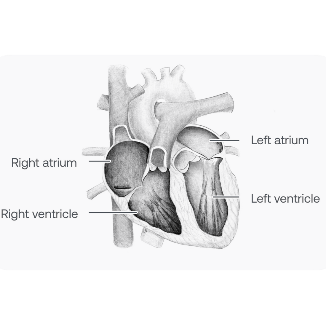
Overview of Cardiac Anatomy

Overview
Mammalian hearts have four chambers — two upper (left and right atria) and two lower (left and right ventricles). The right and left sides of the heart are coordinated, so that the atria contract simultaneously and, after a short delay, the ventricles contract simultaneously. On the right side, deoxygenated blood flows into the heart from the systemic veins, traverses the right atrium, then the right ventricle, and finally is ejected into the lungs. On the left side, oxygenated blood returns from the lungs via the pulmonary veins into the left atrium, then the left ventricle, and then onward to the body.
Heart valves
Healthy heart valves permit the blood in the heart to flow in only one direction and prevent backflow. The atrioventricular (AV) valves are located between the atria and ventricles. These include the tricuspid valve on the right side of the heart and the mitral valve on the left. The semilunar valves are located at the exits of the ventricles and include the pulmonic and aortic valves, which respectively sit at the base of the pulmonary artery and aorta.

Systole
Systole is the period in the cardiac cycle when a cardiac chamber is electrically and mechanically activated and contracting. The atria contract, prompting atrial pressure to increase and blood to be pumped into the ventricles through the AV valves. The ventricles then contract, ventricular pressure increases, and the AV valves close once atrial and ventricular pressures are equalized. Ventricular contraction and pressure growth continues until the pressure of the pulmonary artery (right side) and the aorta (left side) are exceeded. At this point, the semilunar valves open and blood is ejected from the left and right ventricles into the lungs and body, respectively. When systole ends, the cardiac chambers relax, the heart fills with blood from the body, and the cycle repeats.
S1
The closure of the AV valves during systole is heard as the first heart sound or S1. It is difficult to distinguish the mitral and tricuspid valve closure sounds because the valves close at almost exactly the same time. The ejection of blood from the ventricles into the body through the semilunar valves is normally silent.
Heart image showing diastole. Atria and ventricles are relaxed. Semilunar valves are closed and AV valves are open. Blood is flowing from the body into the atria, and passively from the atria into the ventricles.

Diastole
Diastole refers to the time in the cardiac cycle when the cardiac chambers are electrically quiet and relaxed. This relaxation causes the atrial and ventricular chambers to depressurize. When the ventricular pressures fall below the pulmonary and systemic arterial pressures, the semilunar valves close, preventing backflow. When they fall below the atrial pressures, the AV valves open, allowing blood to flow passively into the heart from the body (right side) and the lungs (left side).
S2
The closure of the semilunar valves during diastole is heard as the second heart sound or S2. The aortic and pulmonic components of S2 "split " (are heard separately) because the aortic valve closes slightly before the pulmonic valve. Splitting increases during inspiration, because decreased intrathoracic pressure increases pulmonary arterial capacitance and prolongs right ventricular ejection time. Forced expiration and the Valsalva maneuver increase intrathoracic pressure, decrease pulmonary arterial capacitance, and decrease right ventricular ejection time, so splitting is lost.
Medical Advice Disclaimer
DISCLAIMER: THE CONTENT SET FORTH HEREIN DOES NOT PROVIDE MEDICAL ADVICE OR IS AN ATTEMPT TO PRACTICE MEDICINE
The information, including but not limited to, text, graphics, images, and other material contained on this website are for informational purposes only. No material on this website or document are intended to be a substitute for professional medical education, advice, diagnosis, or treatment.
